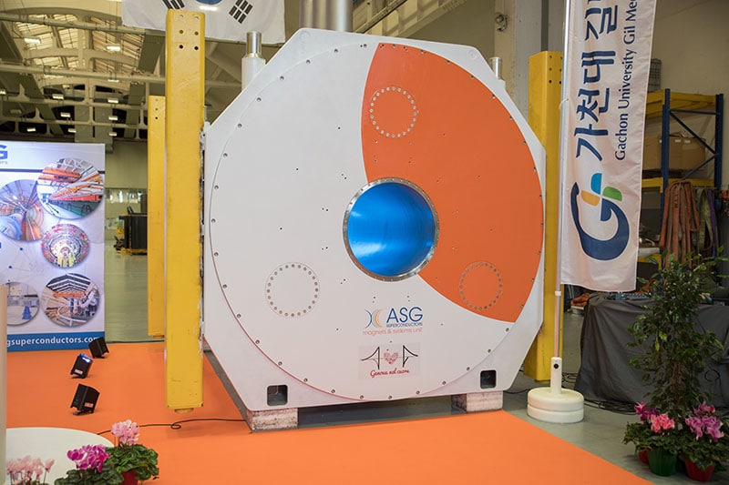
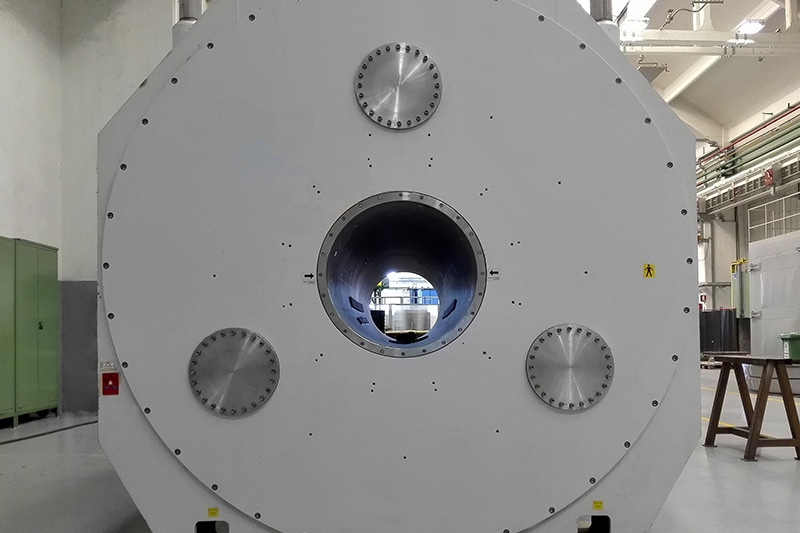
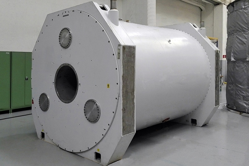
The pelvic floor is a complex 3D support structure for the pelvic organs, which depends on the interplay between the fascia, ligaments and the levator ani muscles. The most commonly used method for prolapse staging is the Pelvic Organ Prolapse Quantification (POP-Q), which is scored in the supine position during physical examination. A recognized limitation of this exam is the scoring of the pelvic organs in the supine position. The objective of this pilot studyis to evaluate the change of prolapse using open standing MRI (MRO) comparing lying, sitting and standing images in women with POP and without (controls).
NEWS
Open Magnetic Resonance Imaging – Thinking Outside the Tunnel
Imaging & Oncology 2017 Open Upright magnetic resonance imaging [...]
Open Upright MRI in the Real World
The spread of open upright MRI scanning is a [...]
The magnets to study the brain
ASG Superconductors has been awarded a contract by Gachon [...]
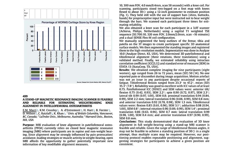
A stand-up magnetic resonance imaging scanner is feasible and reliable for estimating weight bearing knee alignment in patellofemoral osteoarthritis
MRI evaluation of knee alignment in patellofemoral osteo-arthritis (PFOA) [...]
Dynamic MR in patients affected by neurogenical claudication: technique and results from a single-center experience
This study aimed to determine changes in size oflumbar [...]
Findings In Back Pain Patients Referred For Upright MRI
Positional MRI has been developed to provide images of [...]
DESCUBRA MAIS

