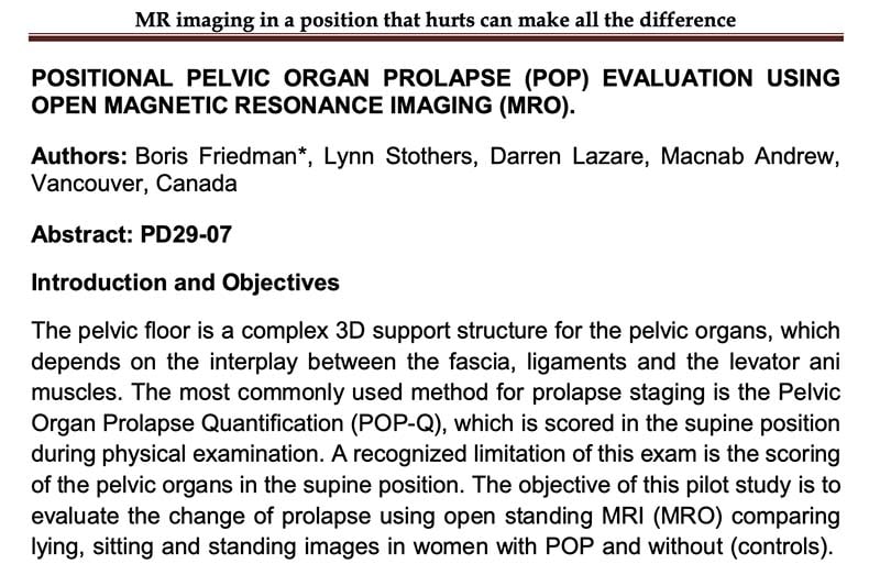
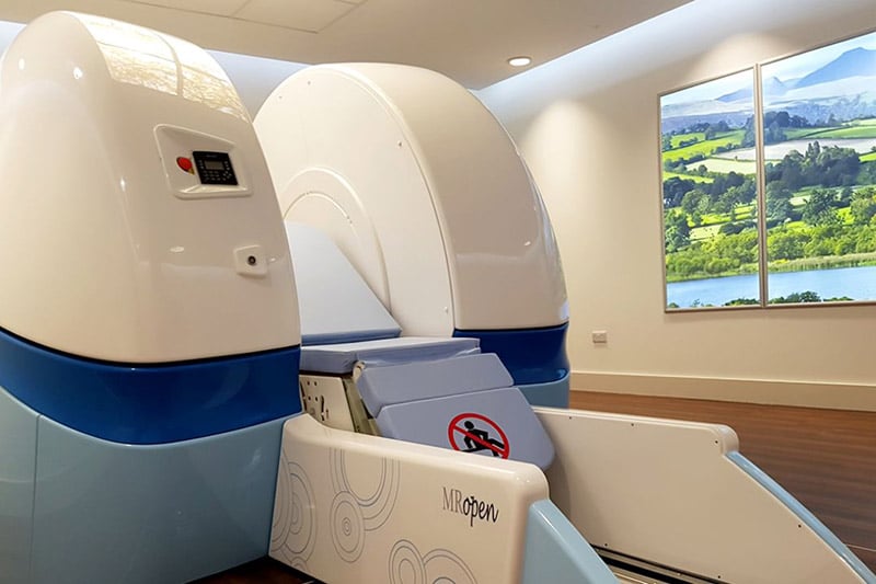
The pelvic floor is a complex 3D support structure for the pelvic organs, which depends on the interplay between the fascia, ligaments and the levator ani muscles. The most commonly used method for prolapse staging is the Pelvic Organ Prolapse Quantification (POP-Q), which is scored in the supine position during physical examination. A recognized limitation of this exam is the scoring of the pelvic organs in the supine position. The objective of this pilot studyis to evaluate the change of prolapse using open standing MRI (MRO) comparing lying, sitting and standing images in women with POP and without (controls).
NEWS
View from the Open MRI in the European Scanning Centre Ltd – Cardiff
The centre situated in Cardiff Gate Business Park contains the [...]
MRI Portfolio optimization – webinar
Penny Gowland, Professor of Physics at the Sir Peter Mansfield [...]
Video: the world’s only OPEN SKY Upright MRI
Discover MROpen EVO features, technology and innovation in the [...]
Penny Gowland – Professor of Physics, Faculty of Science Sir Peter Mansfield Imaging Centre – Nottingham University
Open scanners give a whole new dimension to MRI, which [...]
Alan Breen DC, PhD Professor of MSK Healthcare – AECC University College – Bournemouth (UK)
MROpen can be used both to study the service uses [...]
Patient – Innovative MRI – Orlando (USA)
“.. I have been in many scanners in my life [...]
DISCOVER MORE


