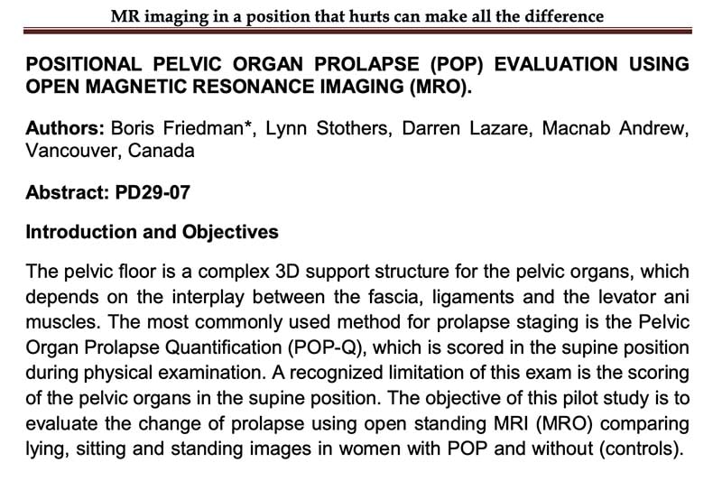
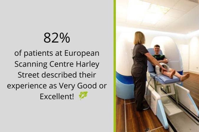
The pelvic floor is a complex 3D support structure for the pelvic organs, which depends on the interplay between the fascia, ligaments and the levator ani muscles. The most commonly used method for prolapse staging is the Pelvic Organ Prolapse Quantification (POP-Q), which is scored in the supine position during physical examination. A recognized limitation of this exam is the scoring of the pelvic organs in the supine position. The objective of this pilot studyis to evaluate the change of prolapse using open standing MRI (MRO) comparing lying, sitting and standing images in women with POP and without (controls).
NEWS
The brand new MROpen Evo website is online!
Introducing the world’s only open MRI scanner. MgB2 helium free [...]
Looking for educational and fun half term activities?
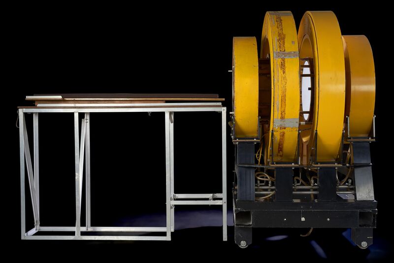
The world's first MRI scanner is currently on display at [...]
Martyn Beckett, Director of Operations, InHealth (UK)
We provide a service to patients who are unable to [...]
Congratulations to the European Scanning Centre
Proud to be part of these results with our unique [...]
MRI Portfolio Optimisation: watch the full webinar!
In this webinar, our Marco Belardinelli (Business Unit Director of [...]
DISCOVER MORE


